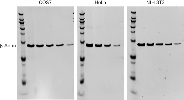
β-Actin Mouse Monoclonal Antibody
Specifications (926-42212)
Size: 100 µL
Storage: -20 °C
Species Cross-Reactivity: human, mouse, rat, monkey, hamster
Target Molecular Weight: 45 kDa
Isotype: Mouse IgG2b
Specificity/Sensitivity: This antibody detects endogenous levels of β-actin protein.
Immunogen: Animals are immunized with a synthetic peptide (KLH-coupled) that corresponds to residues near the amino-terminus of human β-actin.
Storage Buffer: 10 mM HEPES (pH 7.5), 150 mM NaCl, 100 µg/mL BSA, 50% glycerol, and less than 0.02% sodium azide.
Do not aliquot the antibody.
Contains sodium azide.
Recommended Dilution
Western Blotting: 1:1000
In‑Cell Western™ Assay: 1:200
Fixative: Methanol
Applications
This β-actin primary antibody can be used for detection of β-actin by Western blot, or as a normalization antibody when performing two-color detection. Detection of this primary antibody can be achieved with IRDye® Goat Anti-Mouse or IRDye Donkey Anti-Mouse secondary antibodies.
See (licor.com/support) for detailed protocols.
Example Data